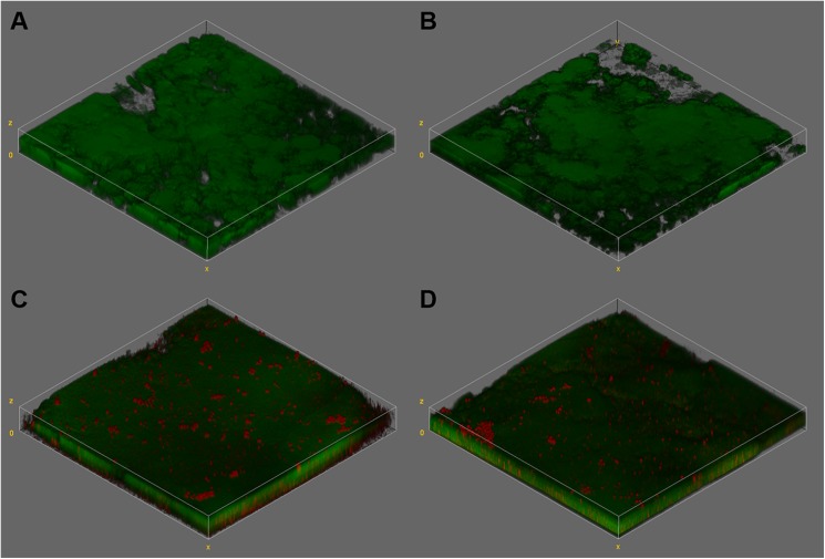
Fig 2.
Tridimensional reconstruction of CLSM images of S. mutans biofilms formed under exposure to glucose + fructose (A and B) or sucrose (C and D) 8x/daily. Images A and C show biofilms visualized at abundance moment, while images B and D at starvation moment. In green, S. mutans cells stained with SYTO 9. In red, extracellular polysaccharides labeled with Alexa Fluor 647—dextran conjugate. Oil immersion objective of 40x (numeric aperture 1.25).

