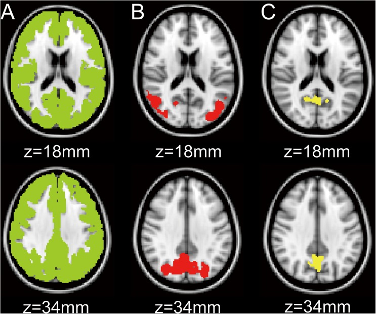
Fig 1. Volumes-of-interest (VOIs) in the MNI space.
A mask of the whole cerebral cortex (A) and the two VOIs (B and C) are displayed on the axial sections of the MNI standard brain. The VOI for B was equal to the significant cluster detected by the voxelwise analysis. The VOI for C was placed on the precuneus, which was sampled from the Harvard-Oxford atlas. MNI: Montreal Neurological Institute.

