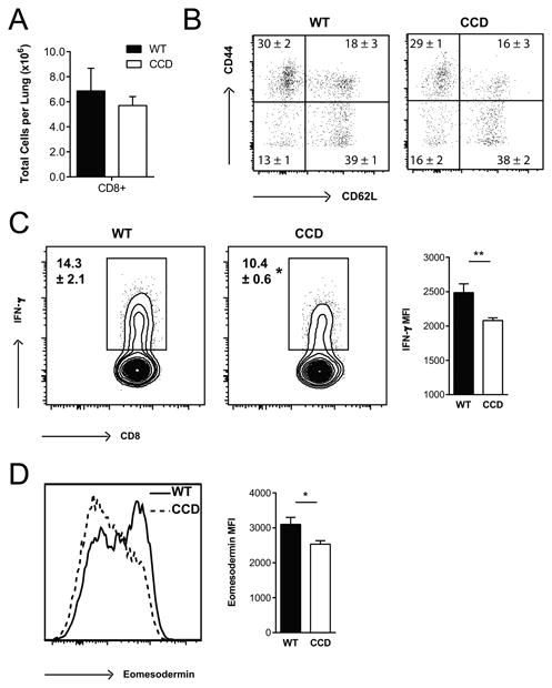
Figure 7. Inhibition of Notch signaling impairs IFN-γ production by CD8+ T cells in the lungs during C. neoformans infection.

Lung leukocytes were isolated from perfused CCD and WT mice at 4 wpi. (A) The total number of CD8+ T cells were quantified and (B) CD44 and CD62L staining was used to assess activation and effector phenotype after gating on Live, CD45+ TCRβ+ CD8+ cells by flow cytometry. (C) Lung leukocytes were stimulated with plate bound anti-CD3 and anti-CD28 antibodies and analyzed for the proportion of CD8+ T cells producing IFN-γ and (D) expression of Eomesodermin was determined by flow cytometry. Flow cytometry plots shown are gated on Live, CD45+, TCRβ+ CD8+ T cells and are representative examples of 1-4 independent experiments. FMO controls were used to set cytokine gates. Frequencies and MFI (geometric mean) data shown are the mean ± SEM with n=5-7/group. *p<0.05, **p<0.01, ***p<0.001
