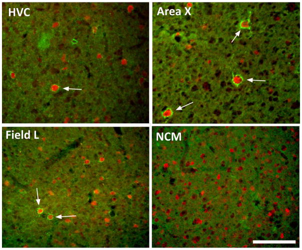
Figure 1.
Representative photomicrographs illustrating the immunostaining for perineuronal nets (PNN; green) and for parvalbumin (red) in two song control nuclei, HVC and Area X and in two auditory brain regions, Field L and NCM of starlings. Arrows point to paravalbumin-immunoreactive cells that were surrounded by PNN. Note the absence of PNN in NCM. Magnification bar = 100 μm.

