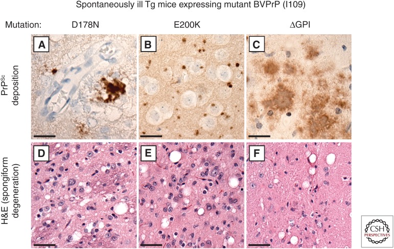
Figure 2.
Prion disease–specific neuropathology in Tg mice expressing mutant bank vole PrP. (A–C) PrPSc deposition, as determined by immunohistochemistry with the antibody HuM-P, and (D–F) spongiform degeneration, as revealed by hematoxylin and eosin (H&E) staining, are apparent in brain sections prepared from spontaneously ill Tg mice expressing D178N-mutant (A,D), E200K-mutant (B,E), or ΔGPI-mutant (C,F) BVPrP(I109). Unique patterns of PrPSc deposition were observed with each mutation: clustered coarse deposits with D178N, small round deposits with E200K, and “plaque-like” deposits with ΔGPI. Scale bars, 20 µm (A–C); 40 µm (D–F).

