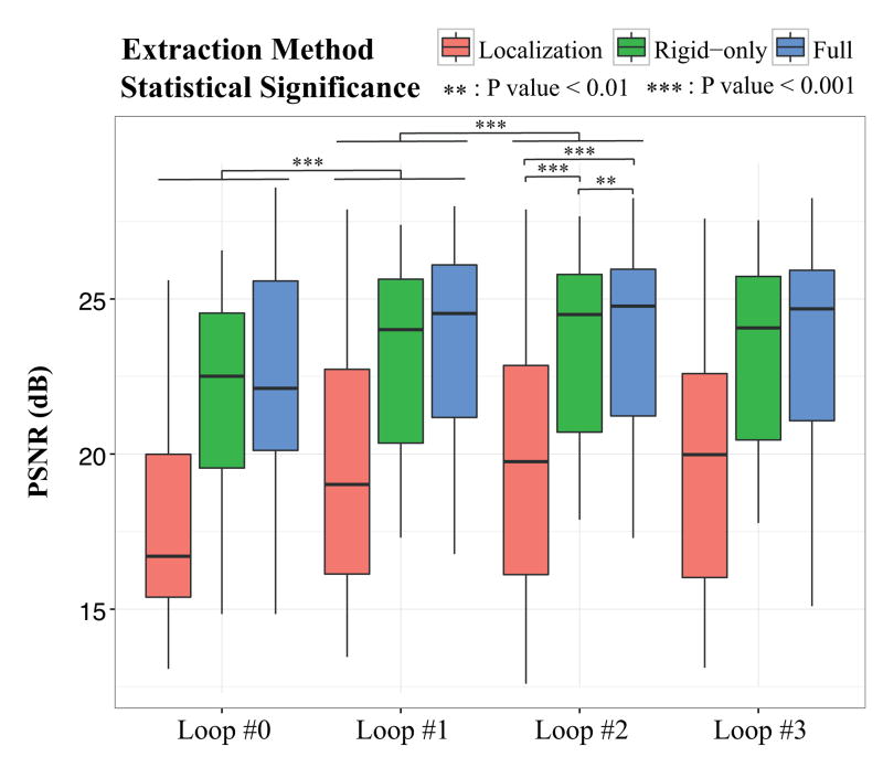
Figure 6.
Influence of brain extraction on image reconstruction quality in terms of Peak-Signal-to-Noise Ratios (PSNR) as reconstruction progresses using (i) only brain localization (Localization-only), (ii) rigid-only slice-to-template registration (Rigid-only) and (iii) the full method (Full). We use as reference the image reconstructed with the help of brain masks manually drawn after brain localization. Loop #L corresponds to the Lth brain mask refinement loop. Loop #0 corresponds to the first image reconstructed using brain masks without any refinement. A two-tailed Wilcoxon test was used for statistical significance evaluation. A significant improvement of the PSNR values can be observed after the first refinement (Loop #1) that becomes not significant after the second refinement (Loop #2). Adopting a rigid-only slice-to-template registration significantly improves the quality of the reconstructed HR image obtained at Loop #2 with an average increase of 3.8dB in the PSNR value. Using the full method allows us to further significantly enhance the quality with an average increase of 4.1dB w.r.t using only brain localization and an average increase of 0.3dB w.r.t using the rigid-only method.

