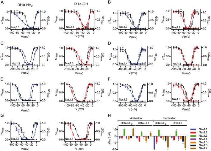
Figure 4.

Modulation of the voltage‐dependence of hNaV channel activation and inactivation gating in the presence of μ‐TRTX‐Df1a. Data (mean ± SEM, n = 5, one cell was considered an independent experiment) for (A) hNaV1.1, (B) hNaV1.2, (C) hNaV1.3, (D) hNaV1.4, (E) hNaV1.5, (F) hNaV1.6 and (G) hNaV1.7 are plotted as G/G max or I/I max. Cells were held at −80 mV. μ‐TRTX‐Df1a C‐terminal amide and acid were applied at respective IC50 concentration for each NaV channel subtype as described in Figure 3 and Supporting Information Table S1. Steady state kinetics were estimated by currents elicited at 5 mV increment steps ranging from −110 to +80 mV. Conductance was calculated using G = I/(V–V rev) in which I, V and V rev are the current value, membrane potential and reverse potential respectively. The voltage‐dependence of fast inactivation was estimated using a double‐pulse protocol where currents were elicited by a 20 ms depolarizing potential of 0 mV following a 500 ms pre‐pulse at potentials ranging from −130 to −10 mV with 10 mV increments. Steady‐state activation and inactivation V50 were determined by the Boltzmann equation. Both C‐terminal acid and amide forms of sDf1a applied at respective IC50 concentrations modified the gating properties of hNaV channels by shifting the voltage‐dependence of activation and steady‐state inactivation to more hyperpolarizing or depolarizing potentials. The ΔV50 was calculated, showing most of the voltage shifts towards more hyperpolarizing potentials, except for hNaV1.3 and hNaV1.7 that had some of these shifts to more hyperpolarizing potentials (H).
