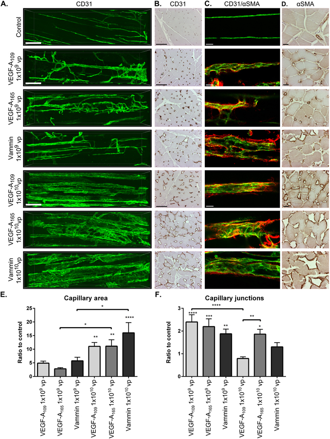
Figure 3.

Both VEGF-A forms and Vammin induce dose and receptor binding profile specific changes in the vascular network (A). Multiphoton microscopy imaging of the blood vessel network by CD31 (green) labelled blood vessels from longitudinal sections of semimebranosus muscle. With 1 × 10⁹ vp dose all tested factors induce sprouting angiogenesis and modest enlargement of capillaries. With 1 × 10¹⁰ vp dose, the normal organization of the blood vessel network is disturbed. VEGF-A109 induces enlarged capillaries that have notably few connections and sprouts. Scalebar 100 µm. (B) CD31 staining of paraffin embedded cross-sections of semimembranosus muscle. Scalebar 100 µm. (C) Confocal microscopy images from CD31 (green) and αSMA (red) double-stained muscles. αSMA positive staining surrounds the enlarged capillaries. Scalebar 25 µm. (D) αSMA staining of the cross-sections shows high coverage of αSMA positive cells in the enlarged capillaries. In control muscles, the staining is mainly seen in the arterioles and venules. Scalebar 25 µm (E) Quantification of the capillary area as ratio to the controls from paraffin embedded CD31 stained cross sections (n = 4–6 per group) (F) Quantification of the amount of capillary junctions as a ratio to the controls from confocal microscopy images of CD31 stained whole-immunomounts (n ≥ 9 images per group). In E to F the data is presented as mean ± SEM. *P < 0.05, **P < 0.01, ***P < 0.001, ****P < 0.0001.
