
Figure 2.
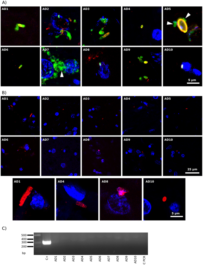
Detection of Borrelia proteins and DNA in ERH samples from ten AD patients. PCR assay of B. burgdorferi DNA. Panel A: ERH sections were immunostained with rabbit polyclonal antibody (1:50) against B. burgdorferi (green) and rat polyclonal antibody (1:20) against T. viride (red). Panel B: ERH sections were immunostained with mouse monoclonal antibody (1:10) against B. burgdorferi (green) and rabbit polyclonal antibody (1:100 dilution) against C. albicans (red). DAPI staining of nuclei appears in blue. Scale bar as shown in the figure. Panel C: PCR analysis of Borrelia DNA in frozen brain tissue from ten AD patients. Nested PCR analysis of ten ERH samples using Borr primers to amplify flagellin gene. The primers employed were Borr FE–Borr RE for the first PCR and primers Borr FI–Borr RI for the second PCR. As positive control, DNA extracted from B. burgdorferi was used. Control PCR: PCR without DNA. DNA markers are indicated on the left.