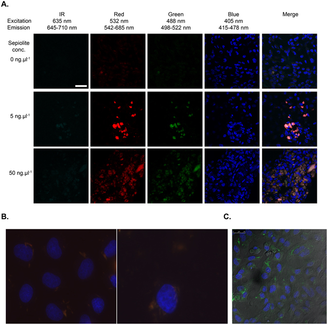
Figure 1.

Laser confocal microscopy images of sepiolite fibers (0, 5 and 50 ng·μl−1) in V79 cells. (A) Channels: IR (infrared), red, green and blue, at given excitation and emission wavelengths. Sepiolite has a natural fluorescence in the green and red ranges and is not fluorescent in blue or infrared. The blue fluorescence represents the Dako-DAPI staining of the cell nuclei. In merged images, we confirmed that sepiolite fibers were uptaken by the cells. IR: infrared. The sepiolite concentration is indicated on the figure. The wavelengths of excitation (Exc) and fluorescence emission (Emis) are indicated. (B) Magnified fluorescence microscopy images of sepiolite fibers in V79 cells. Nucleus in blue (DAPI staining), sepiolite fibers in orange/red. (C) Phase contrast in confocal analysis visualizing cell contour and the presence of sepiolite (green) inside the cell.
