Figure 1.

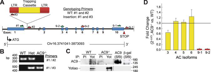
Design and verification of the AC9−/− mouse model. (A) Schematic of AC9 gene-trap strategy and genotyping primers. Intron distances are not drawn to scale. (B) PCR analysis of genotyping: lanes 1, 2, and 3 represent wild-type (WT), AC9+/− (Het) and AC9−/−, respectively. (C) AC9 protein levels were detected by immunoprecipitation of pre-immune (PI) or Yotiao complexes from WT and AC9 KO heart extracts followed by western blotting (WB) with anti-AC9 antibody. AC9 protein is not detectable in total heart extracts by WB (n = 5). (D) Real time PCR of cardiac AC isoforms in AC9−/− heart, normalized to WT expression levels (n = 3; mice 1 month of age). Loss of AC9 mRNA was confirmed with primer sets 9-1 and 9-2. Full-length WBs are presented in Supplementary Fig. S6.
