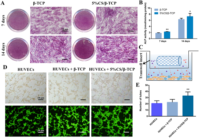
Figure 3.

5%CS/β-TCP scaffolds stimulate hBMSC osteogenesis and HUVEC angiogenesis in vitro. (A) ALP staining in hBMSCs cultured for 7 and 14 days with β-TCP scaffolds or 5%CS/β-TCP scaffolds in transwell inserts. (B) 5%CS/β-TCP scaffolds stimulated ALP activity at 7 days and 14 days in comparison to pure β-TCP. (C) Schematic representation of transwell experiments. Scaffolds in Transwell inserts (upper chamber), cells in the lower chamber. (D) Tube formation by HUVECs, as observed by light microscopy (top) and Calcein AM staining (bottom), after 4 h on Matrigel with β-TCP or 5%CS/β-TCP scaffolds in transwell inserts. (E) 5%CS/β-TCP scaffolds enhanced tube formation in comparison to β-TCP scaffolds. *p < 0.05; **p < 0.01. Scale bar represents 10 μm.
