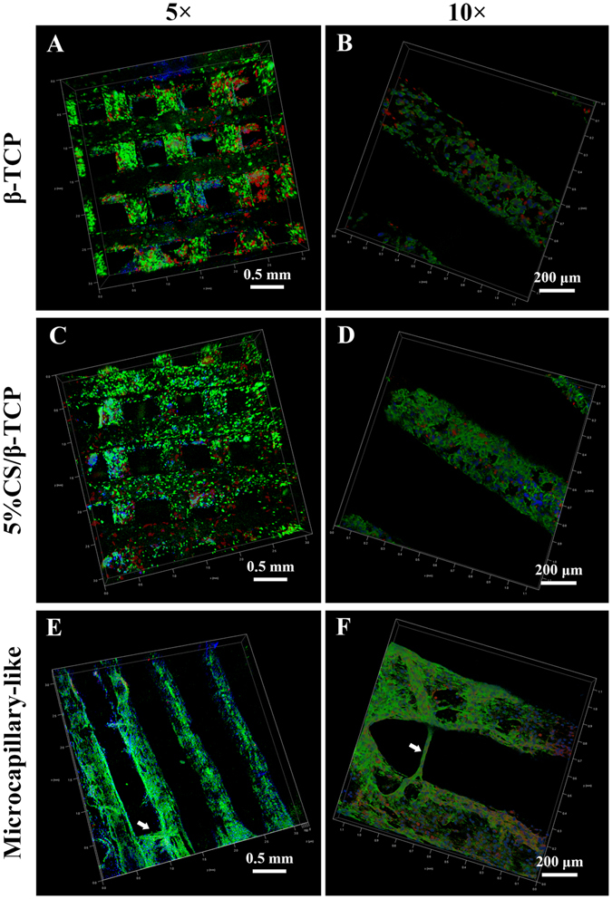
Figure 5.

Confocal laser scanning microscopy of co-cultured cells on β-TCP and 5%CS/β-TCP scaffolds. Confocal images of HUVECs co-cultured with hBMSCs on (A,B) β-TCP and (C,D) 5%CS/β-TCP scaffolds for 3 days. Viable cells were stained with FITC (green), nuclei were stained with DAPI (blue), and hBMSCs were tracked with CM-Dil (red). (E,F) Confocal images of HUVECs co-cultured with hBMSCs labeled with CM-Dil (red) on 5%CS/β-TCP scaffolds for 10 days. Endothelial cells were stained with CD31 (green), and nuclei were stained with DAPI (blue). White arrows mark CD31-positive microcapillary-like structures.
