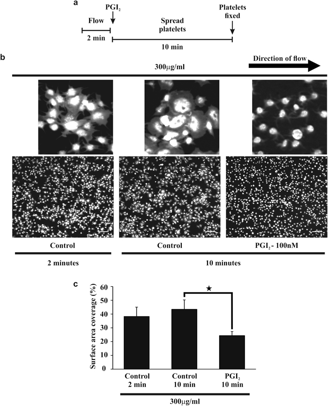
Figure 1.

Post perfusion of 100 nM PGI2 induces embolisation of performed thrombi on fibrinogen. Whole blood, stained with 10 μM DiOC6 was flowed over fibrinogen (300 μg/ml) coated slides for 2 minutes at a shear rate of 1000 s−1, to enable the formation of thrombi. After 2 minutes either tyrodes alone or tyrodes containing 100 nM PGI2 were perfused over the preformed thrombi for 10 minutes at 1000 s−1. (a) Schematic of the experimental condition of the flow. (b) Representative images of the thrombi observed under these different experimental conditions. Scale bar is 20 μm. The insert shows an enlarged image of their respective images with a scale bar of 5 μm. (c) The surface area of the thrombi at 2 minutes of flow and after 10 minutes of perfusion with 100 nM PGI2. Thrombi were fixed with 4% paraformaldehyde, before restaining with 10 μM DiOC6 overnight. Thrombi were then imaged using flourescent microscopy, and analysed using ImageJ to obtain the surface area. Analysis completed from n = 3 experiments. p < 0.05.
