
Figure 1.
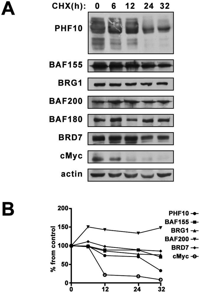
(A) The stability of PBAF subunits in human cells. HEK293 were treated with 20 µg/ml cycloheximide (CHX) for the time points indicated above the panels. The equal amounts of lysates of control (0 h) and CHX-treated HEK293 were then analyzed by Western blotting. β-Actin was used as loading control. (B) The intensity of bands on Western blot was quantified using ImageJ software by densitometry as described in Methods section. The images in Fig. 1(A) is cropped.