
Figure 2.
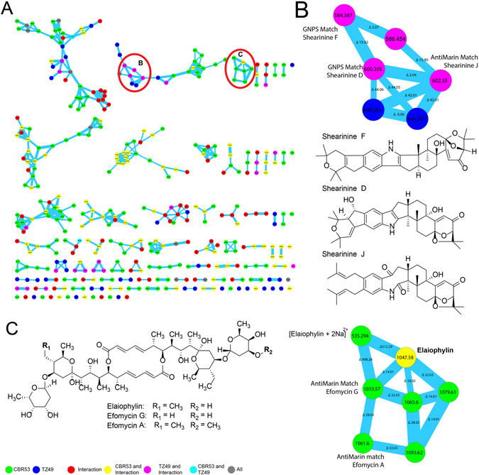
Molecular network from the extract of the bacterium Streptomyces CBR53 with the identification of the elaiophylin macrolide. (A) Molecular network after filter blanks, colors indicate producers of the nodes: Streptomyces CBR53 green; Escvopsis TZ49 blue, Streptomyces sp. vs Escovopsis sp. (CBR53 vs TZ49) red. Nodes found in more than one producers are represented as combination of their colors: Streptomyces CBR53, Streptomyces sp. vs Escovopsis sp. (CBR53 vs TZ49) yellow; Escovopsis TZ49, Streptomyces sp. vs Escovopsis sp (CBR53 vs TZ49) pink; Streptomyces CBR53, Escvopsis TZ49 cian. (B) Enlarged cluster of shearinine derivatives indicating their location in the sub-network, chemical structures of shearinine D, shearinine F and shearinine J. (C) Chemical structures of elaiophylin, efomycin G and efomycin A macrolides dereplicated using GNPS and AntiMarin, enlarged cluster denoting their location in the cluster.