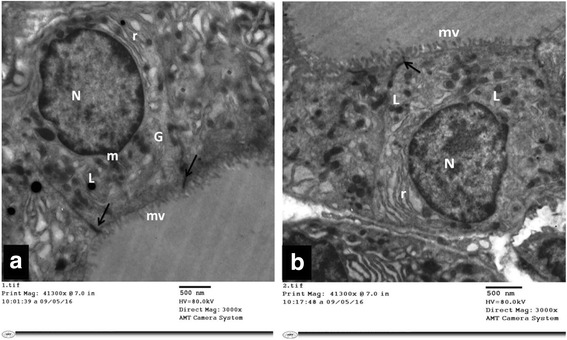
Fig. 10.

a & b: Electron micrographs of thyroid follicular cells of TBT + GTE group (Group III). It is showing cuboidal cells with a well-developed microvillous (mv) border. The nucleus (N) is euchromatic and rounded. The cytoplasm shows normal to mildly dilated rough endoplasmic reticulum (r), lysosomes (L), mitochondria (m) and Golgi apparatus (G). An intact tight junction (↑) is also noticed
