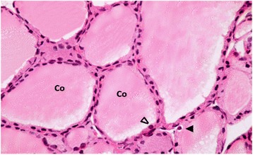
Fig. 4.

A Photomicrograph of a section in thyroid gland of a TBT-treated rat that received green tea (group III). It is showing moderately vacuolated colloid (Co) filling the follicular lumen. Most of the follicular cells appear with oval to rounded nuclei (∆) and normal cytoplasm, while others still reveal mild vacuolated cytoplasm (▲). H&E stain Mic.Mag. X 400
