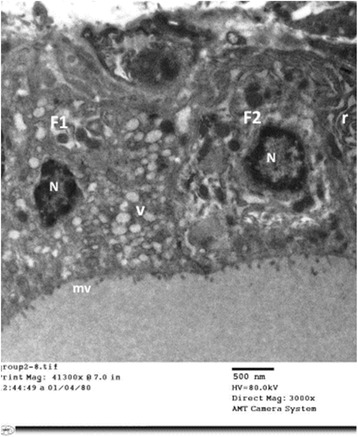
Fig. 7.

An electron micrograph of thyroid follicular cells of TBT-treated rat (Group II). It is showing two cuboidal cells (F1&F2). The microvilli (mv) are short and blunt. The nuclei (N) are small and irregular with peripheral clumping of heterochromatin. F1 cell exhibits numerous vesicles (V). F2 cell depicts dilated profiles of rough endoplasmic reticulum (r)
