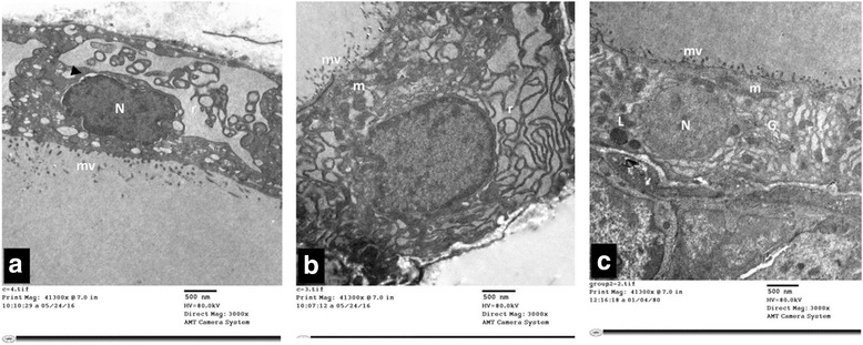
Fig. 8.

a, b & c: Electron micrographs of thyroid follicular cells of TBT-treated rat (Group II). It reveals cuboidal follicular cells with microvillous border (mv). The cytoplasm is filled with extensively dilated rough endoplasmic reticulum (r) that filled with flocculent material. Photo a shows irregular nucleus (N) with dilated perinuclear cisterna (▲). Photo b depicts mitochondria (m) with disrupted cristae. A large Golgi complex (G) is encountered in photo (c). Notice: (L); lysosomes
