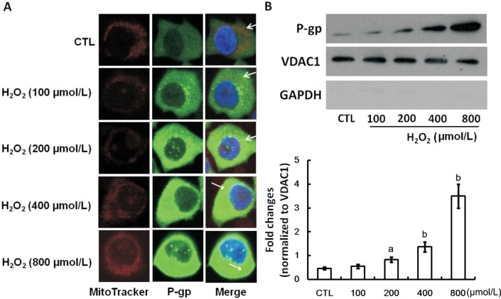
Figure 2. Mitochondrial expression and location of P-gp in untreated and H2O2-incubated D407 cells.
A: Representative confocal micrographs of D407 monolayers showed that D407 cells have exact overlapping P-gp staining (green) and mitochondria staining (red), indicating colocalization of P-gp and mitochondria (as shown in white arrowhead); B: A representative Western blot picture showed the alterations of mitochondrial P-gp in translational level in D407 cells after exposing to varying concentration of H2O2 for 24h. VDAC1 was used as the internal loading control of mitochondrial proteins and GAPDH was used as a cytoplasm marker. The relative protein expression was expressed as the gray value ratio of target protein to respective VDCA1. The average expression of each protein was calculated from three independent experiments. aP<0.05, bP<0.01 versus CTL for one-way ANOVA. CTL: Untreated cells.

