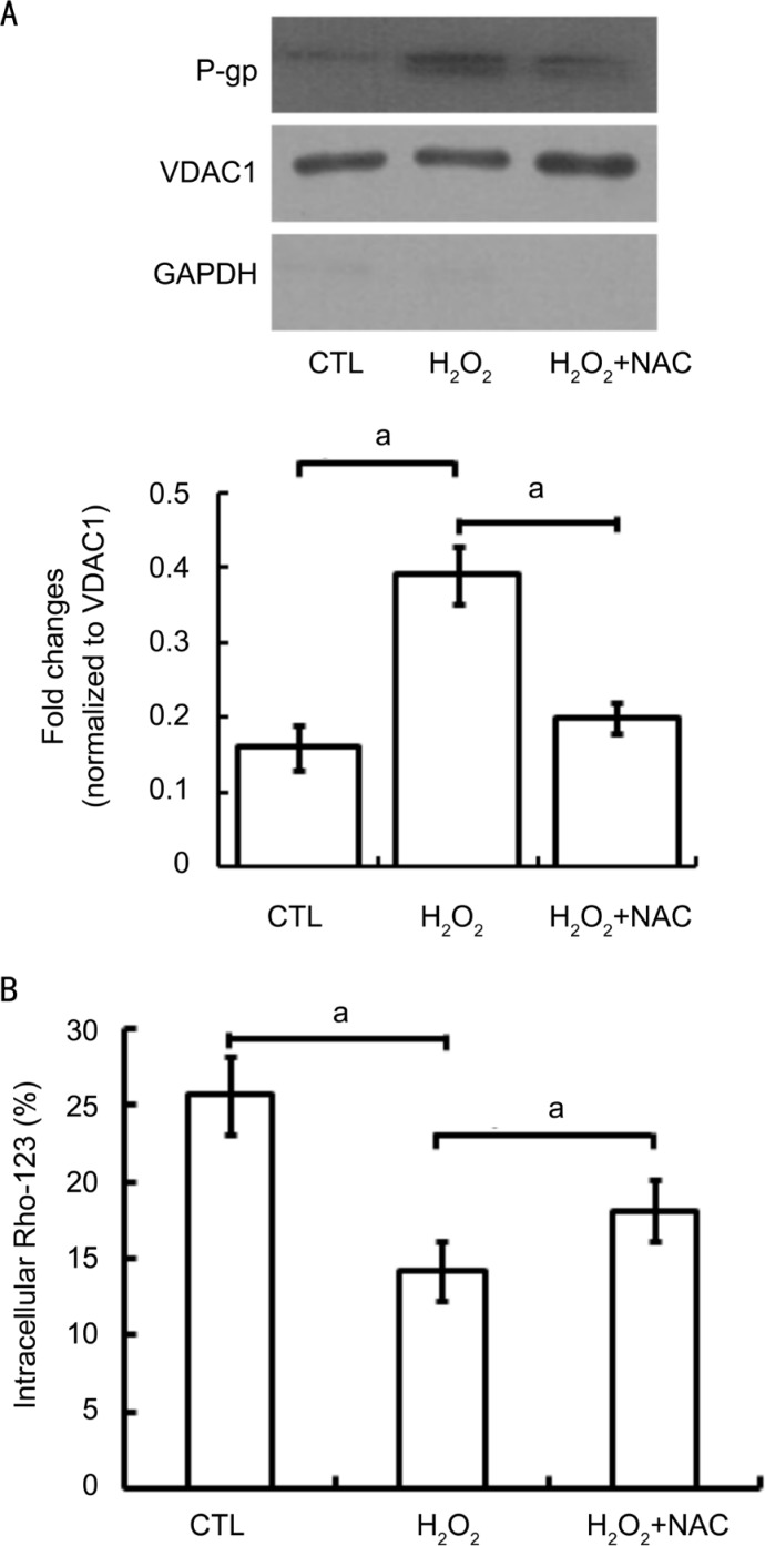
Figure 5. Effects of antioxidants on the functional expression of P-gp in mitochondria of D407 cells.
D407 cells were treated with 200 µmol/L H2O2 alone or in combination with 10 mmol/L NAC (30min prior to H2O2 treatment) for up to 24h. Untreated cells were set as the control (CTL). The mitochondrial expression of P-gp was detected by Western blot and the activity of P-gp was determined by measuring intracellular Rho-123 accumulations by flow cytometry. A: A representative Western blot image shows the mitochondrial expression of P-gp. VDAC1 was used as the internal loading control of mitochondrial proteins and GAPDH was used as a cytoplasm marker; B: Quantitative densitometric analysis of the mean fluorescence intensity of intracellular Rho-123 following different treatment was performed in three independent experiments. aP<0.05 versus CTL for one-way ANOVA.

