Figure 2.
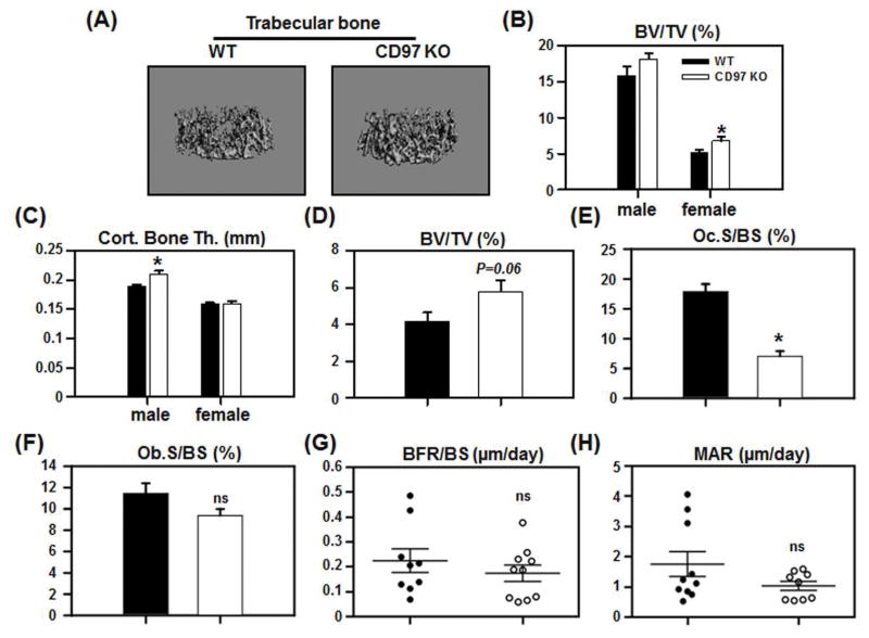
CD97 KO mice had increased bone mass with decreased osteoclast number. (A–C) The femurs from WT and CD97 KO mice were analyzed by micro-CT. A) Representative images of trabecular bone from WT and CD97 KO female mice. B) Trabecular bone volume (BV/TV); C) Cortical bone thickness. (D–F) Static histomorphometric analysis. The femurs from WT and CD97 KO female mice were sectioned, stained for H&E and TRAP and were analyzed by Osteomeasure. D) Trabecular bone volume (BV/TV); E) osteoclast surface (Oc.S.BS); F) osteoblast surface (Ob.S/BS). (G–H) Dynamic histomorphometric analysis. Mice were injected with calcein 9 and 2 days prior to sacrifice. Frozen sections from the femurs of WT and CD97KO mice were evaluated. (G) Bone formation rate per bone surface; (H) Mineral apposition rate. Values represent mean SEM. N=6–11. *, Significant effect of CD97 KO mice, p<0.05. ns: not significant.

