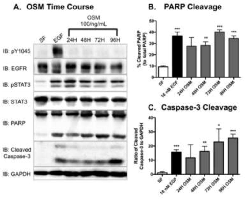
Figure 4. EGFR-independent activation of STAT3 promotes apoptosis in MDA-MB-468 cells.

A. Serum-starved MDA-MB-468 cells were stimulated with 100 ng/mL of Oncostatin M (OSM) for the indicated times. Control cells were exposed to serum free DMEM (SF) and or 16 nM EGF for 24 hours. After harvesting, the cell lysates (40 μg) were resolved by SDS PAGE, and were immunoblotted for the indicated proteins. Densitometry quantification of western blot data of cleaved PARP (B.) and cleaved Caspase-3 (C.). The cleaved PARP band intensities were normalized to and plotted as a percentage of total PARP bands. Cleaved Caspase-3 bands were quantified to total protein for each respective sample (GAPDH). Data are expressed as the average ± SEM (n=3).
