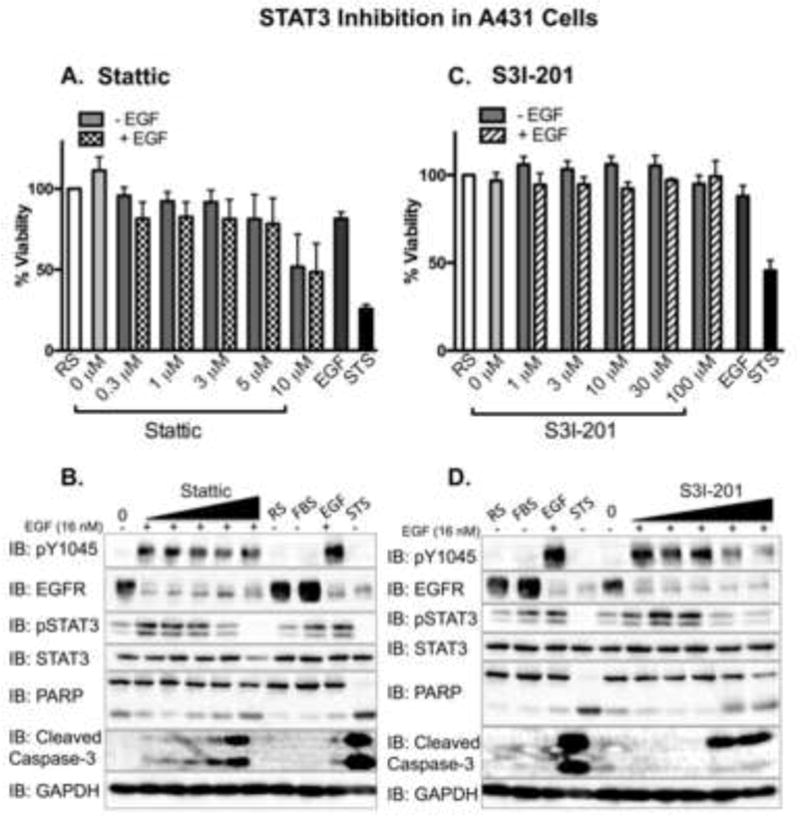
Figure 6. STAT3 inhibitors attenuate EGFR activity in A431 cells.

A431 cells, plated and treated in parallel, were assessed for cell viability using an Alamar Blue assay. Data from A. Stattic or B. S31-201 treated cells are expressed as the average ± SEM (n=3) relative to cells incubated in 1.25% FBS in DMEM (reduced serum – RS). Serum-starved A431 cells were pre-treated for 1 hour with 0 (0.025% DMSO), 0.3, 1, 3, 5, or 10 μM Stattic (IC50 = 5.1 μM; panel A.), or with 0 (0.5% DMSO), 1, 3, 10, 30, and 100 μM S3I-201 (IC50 = 86 μM; panel C.), followed by the addition of 16 nM EGF (+) for 24 hours. Control cells were incubated with reduced serum media alone (RS), media containing 10% fetal bovine serum (FBS), or 16 nM EGF (EGF) for 24 hours. As a positive control cells were treated with 1 μM staurosporine (STS) for 4 hours. Cell lysates were prepared, 40 μg were resolved by SDS-PAGE, and immunoblotted for the indicated proteins. Fifteen μg of the STS sample were used to keep the immunoblot analysis in the dynamic range. Shown is a representative experimental replicate of three.
