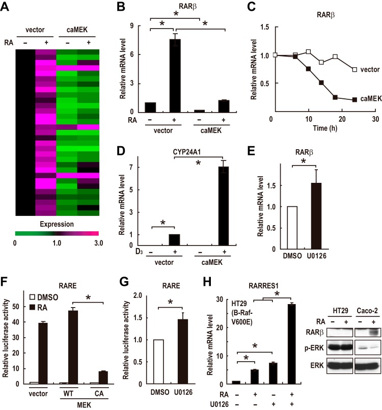
FIG 6.
ERK activation suppresses transcriptional activity of RAR. (A) Expression profiles of the RAR target genes in CRC cells are shown. Cells were infected with an adenovirus expressing constitutively active MEK (caMEK) or a control virus (vector) 12 h before treatment with DMSO or RA (100 nM). Cells were incubated for 24 h, and total RNA was prepared and subjected to RT-PCR analysis. Each horizontal line displays the expression data for one gene, where the ratio of the mRNA level to its level in the control cells, which were infected with a control virus (vector) and treated with DMSO, is represented by color according to the color scale. (B) Caco-2 cells were treated as described for panel A. The mRNA level of RARβ was analyzed by RT-PCR. (C) The expression levels of the RARβ mRNA in confluent Caco-2 cells infected with an adenovirus expressing constitutively active MEK (caMEK) or a control virus (vector) were evaluated by RT-PCR. (D) Confluent Caco-2 cells were infected with an adenovirus expressing constitutively active MEK (caMEK) or a control virus (vector). At 12 h postinfection, cells were treated with DMSO or 1,25-(OH)2-vitamin D3 (100 nM) for 24 h. Total RNA was prepared and analyzed by RT-PCR. (E) The expression levels of the RARβ mRNA in preconfluent Caco-2 cells treated with U0126 (10 μM) or DMSO vehicle for 24 h were analyzed by RT-PCR. (F and G) Transcriptional activation of a reporter plasmid expressing luciferase under the control of the RA-responsive elements (RARE3-Luc) was evaluated in Caco-2 cells transfected with empty vectors or with expression plasmids for wild-type (WT) or constitutively active (CA) MEK. Cells were treated with 100 nM RA for 12 h (F) or with 10 μM U0126 for 24 h (G) prior to the measurements. (H) The expression levels of the RARRES1 mRNA in HT29 cells treated with 1 μM RA and/or 10 μM U0126 for 72 h were analyzed by RT-PCR (left). HT29 and Caco-2 cells were treated with DMSO or 100 nM RA for 24 h, and cell lysates were prepared and analyzed by immunoblotting (right). Values in panels B and D to H are means ± standard errors of the means (n = 3). *, P < 0.05.

