Fig. 3.

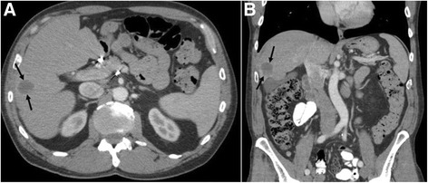
a Axial and b coronal post-contrast CT images through the abdomen during PD-1 therapy again demonstrate the index metastatic lesion (arrows) within the right hepatic lobe with post-treatment changes and marked decrease in size

a Axial and b coronal post-contrast CT images through the abdomen during PD-1 therapy again demonstrate the index metastatic lesion (arrows) within the right hepatic lobe with post-treatment changes and marked decrease in size