
Fig. 3.
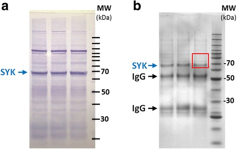
Enrichment of SYK for relative quantitative measurement of phosphorylation. (a), In triplicate samples, 25 μg of WEHI cell extract was separated by SDS-PAGE and SYK mobility was visualized by colorimetric western blotting. A major band close to the predicted molecular weight of SYK (71 kDa) is indicated by a blue arrow. Location of a molecular weight (MW) ladder is indicated on the right, which occurs in 10 kDa increments. (b), SYK was immunoprecipitated as described in the Material & Methods. Samples were separated by SDS-PAGE and the gel stained with Coomassie Blue. Major bands corresponding to SYK as well as heavy and light IgG chains are indicated. For multiple reaction monitoring (MRM) experiments, areas of the gel corresponding to the red box were excised