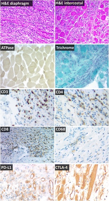
Fig. 2.

Representative immunohistochemistry of inspiratory muscles at autopsy, 66 days after tremelimumab-durvalumab treatment. Hematoxylin and eosin (H&E) sections show inflammatory myopathy in the diaphragm and intercostal muscles without rimmed vacuoles and without perifascicular atrophy, consistent with polymyositis. A mononuclear infiltrate is present which invades otherwise normal myofibers and completely effaces the background muscle fiber architecture in some areas. ATPase shows intense staining in small fibers compared to surrounding lighter, normal sized fibers in preserved areas of muscle, indicative of type II fiber atrophy. Trichrome shows mildly increased connective tissue, but shows no rimmed vacuoles, rods, or other inclusions. T cell co-receptor staining (CD3, CD4, CD8) revealed a mixed T-cell infiltrate which often completely effaced the myofascicular architecture. CD68 highlights necrotic myofibers scattered within the larger inflammatory infiltrate. PD-L1 expression was observed in blood vessels of dying muscle. Weak CTLA-4 expression was detected in necrotic myofibers. All images are 100× magnification
