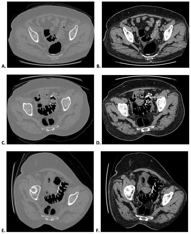
Figure 1.


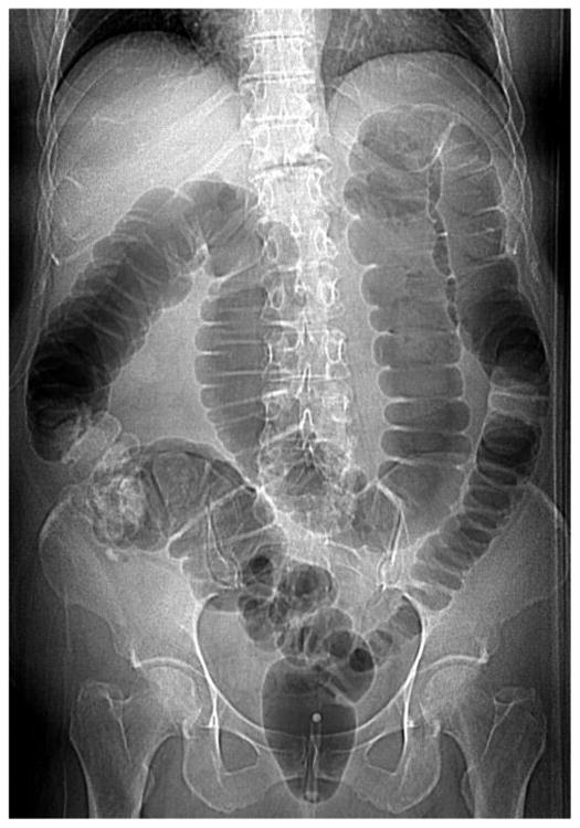
Decubitus position obtained for nondiagnostic sigmoid distention on both supine and prone series at CTC screening in an asymptomatic 60-year-old woman.
Supine CTC images with polyp (A) and soft tissue (B) windowing show complete collapse of a long segment of sigmoid colon. Prone CTC images (C and D) show somewhat improved sigmoid distention but with persistent focal areas of collapse. Given these findings, the CT technologist obtained a third series in the right lateral decubitus position (E and F), which demonstrate good sigmoid distention, leading to overall diagnostic adequacy. Note how the segmental collapse is not apparent on the supine scout view (G), which is why we require online review of the 2D images in cine mode at the CT console by the. The majority of cases of inadequate distention involve the sigmoid colon.
