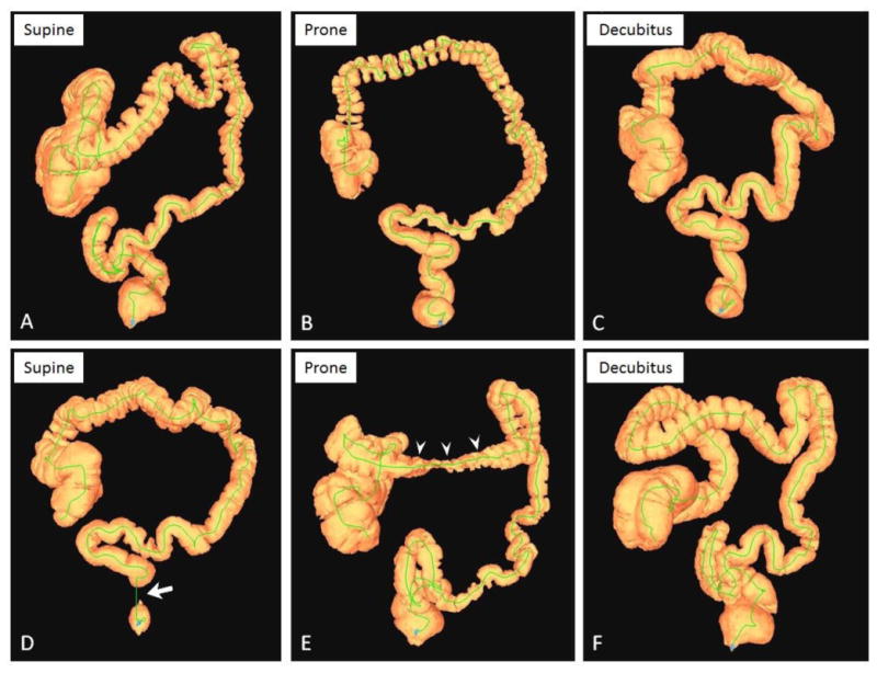
Figure 1.

3D colon maps from CTC in two patients showing typical distention patterns.
A-C, CTC 3D colon maps from a 67-year-old man show intermediate/adequate distention in the supine position (A, colonic gas volume = 1429 ml), poor/inadequate distention in the prone position (B, gas volume = 973 ml), and optimal distention in the right lateral decubitus position (C, volume = 1963 ml). D-F, CTC 3D maps from a 51-year-old woman in the supine (D), prone (E), and decubitus (F) positions with colonic gas volumes of 1835 ml, 1639 ml, and 2452 ml, respectively. Note typical areas of inadequate distention in the rectum on supine (D, arrow) and transverse colon on prone (E, arrowheads), both of which are much better distended on the decubitus series (F).
