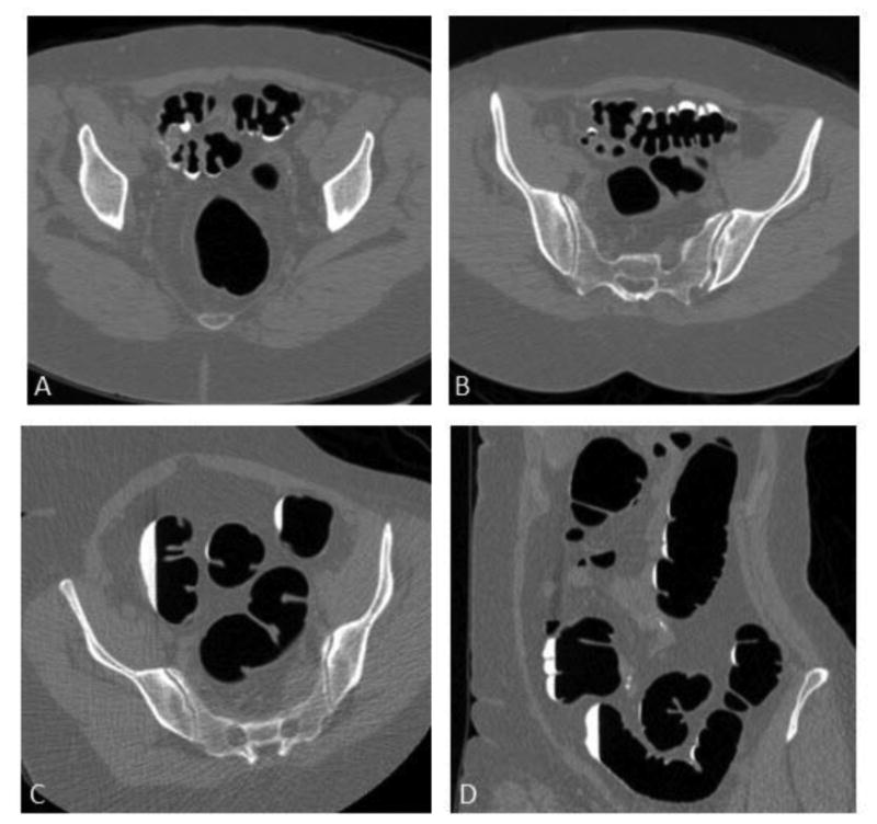
Figure 2.

2D images from CTC examination in 65-year-old woman.
Transverse 2D CTC images from supine (A), prone (B), and right lateral decubitus (C) series show improved distention on the decubitus series. Coronal 2D decubitus image (D) also depicts excellent distention. Resolution of luminal narrowing and crowding of colonic folds greatly simplifies evaluation on both 2D and 3D views. Colonic gas volumes in supine, prone, and decubitus were 1388 ml, 1382 ml, and 2141 ml, respectively.
