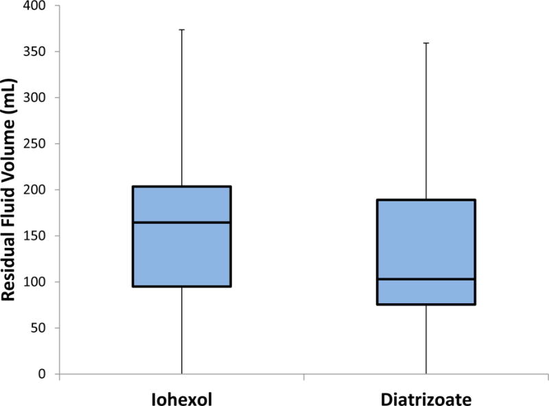
Figure 2.

Standard box-and-whisker plots of residual colonic fluid volumes. Results are clinically comparable. Boxes include all measurements in the 2nd and 3rd quartiles with the line inside the box denoting the median value. Whiskers indicate 1.5 × the inter-quartile range (portions of the lower whiskers are cropped because they extend below 0).
