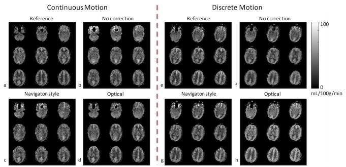
Figure 3.
CBF maps from the first volunteer’s scan in the presence of continuous and discrete motion. Without correction, the CBF maps contain significant blurring, especially in the frontal part of the brain where the pixel-wise displacement is the largest (b,f). With navigator-style correction, the CBF maps still have residual artifacts due to the delay in correction (c,g). Images corrected with optical tracking (d,h) are very similar to images with no motion (a,e).

