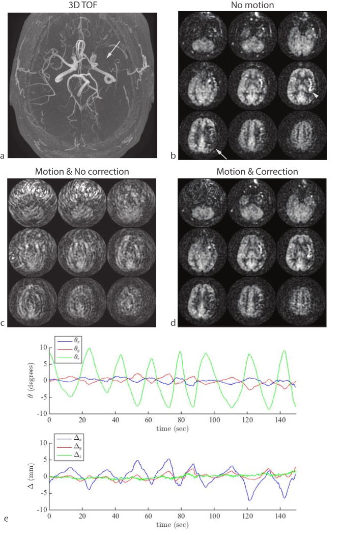
Figure 8.
Result of continuous shaking motion in a patient with chronic steno-occlusive disease. (a) An axial MIP from a 3D TOF MRA shows absence of blood flow in the left middle cerebral artery (arrow). The patient was asymptomatic and had sufficient collateral supply but the delayed arrival of the arterial spin label can be seen as a region of decreased ASL signal in the left M1 territory tissue (b, arrow) and hyper-intense arterial signal in the left insula region (b, arrowhead). This signal abnormality was masked by motion (c), but was visible in a second measurement that included optical motion correction (d). The motion plot from the optical-corrected scan is shown in (e).

