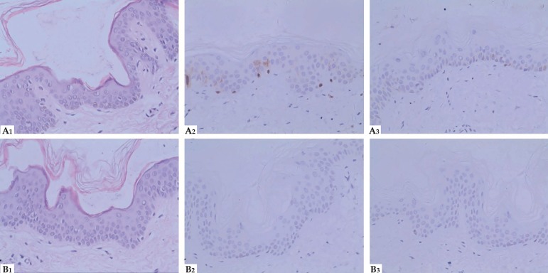
Figure 3.
Histopathological and immunohistochemical features( Hematoxylin & eosin x400). HE staining (A1), Immunohistochemical staining for S100 (A2) and HMB45 (A3) respectively from hyperpigmentation; HE staining (B1), Immunohistochemical staining for S100 (B2) and HMB45 (B3) respectively from hypopigmentation

