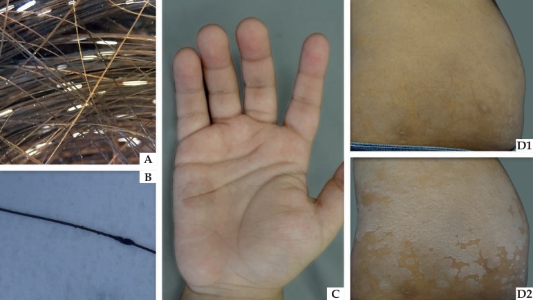
Figure 1.
Clinical appearance of actual superficial mycoses. (A) White piedra: whitish nodule attached to the hair shaft. (B) Black piedra: darkened nodule attached to the hair shaft. (C) Tinea nigra: brownish macula on children’s palms. (D) Pityriasis versicolor: scattered maculas on the abdomen (D1), which become more evident after skin stretching (Zireli’s sign – D2)

