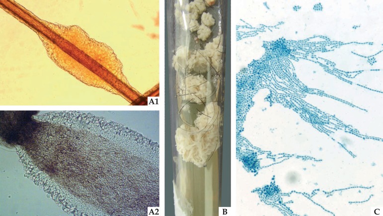
Figure 2.
Mycological examinations of white piedra: (A1) Optical microscopy (x40) offering a detailed illustration of the light color nodule attached to the pillar shaft. (A2) Optical microscopy (x100) illustrates the yeasts the make up the structure on the edge of the nodule. (B) Culture Mycosel medium (Difco, USA) with yeast-like colony, with the cerebriform filamentous appearance. (C) Microgrowth demonstrates yeasts with blasto-arthrospores, typical of Trichosporon sp

