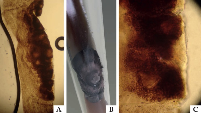
Figure 3.
Mycological examinations of black piedra: (A) Optical microscopy (x40) offering a detailed illustration of the dark nodule attached to the pillar shaft. (B) Culture Mycosel medium (Difco, USA) with dematiaceous colony. (C) Optical microscopy (x100) identifying the ascus, round structures typical of parasitism caused by Piedraia hortae

