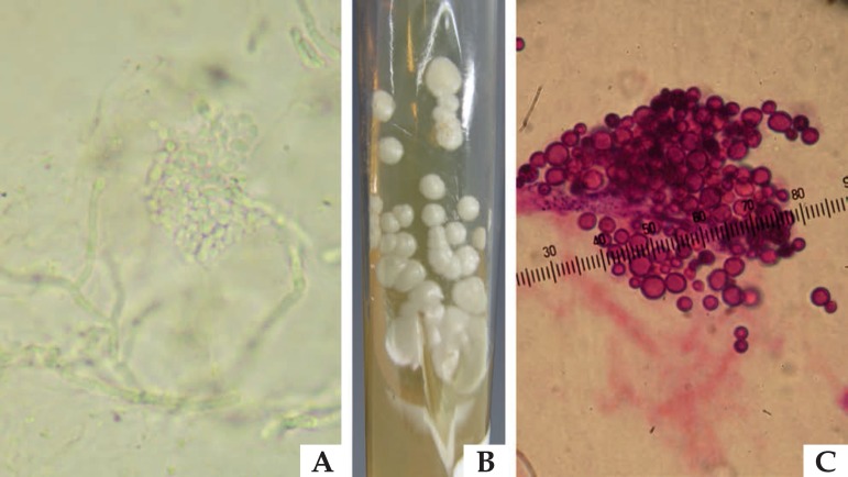
Figure 5.
Mycological examinations for pityriasis versicolor: (A) Direct mycological examination of a sample collected through skin lesion scraping, clarified with KOH 10%, illustrating yeasts grouped in a “grape bunch” format, and of short and thick pseudo-hyphae. (B) Sabouraud agar culture, enriched with olive oil, with beige yeast-like colony. (C) Yeasts grouped with short base single budding, with “bowling pin” appearance, stained by the hematoxylin eosin method, typical of Malassezia sp microgrowth

