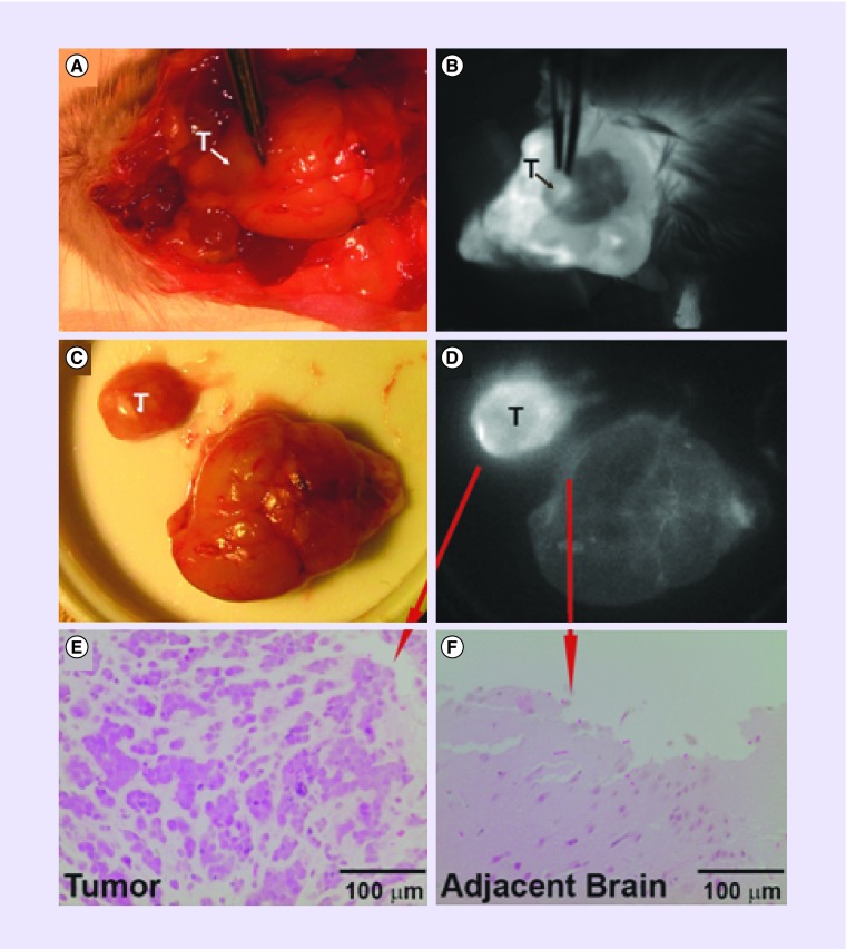
Figure 4. . CLR1502 fluorescence imaging of a patient-derived glioblastoma stem cell orthotropic xenograft.
(A & B) White light and fluorescence imaging of glioblastoma stem cell xenograft and (C & D) after resection. (E & F) Verification of tumor and normal brain by histology.
Reprinted with permission from [13] © Wolters Kluwer Health, Inc.

