. 2014 Oct 10;6(1):301–307. doi: 10.1039/c4sc02600d

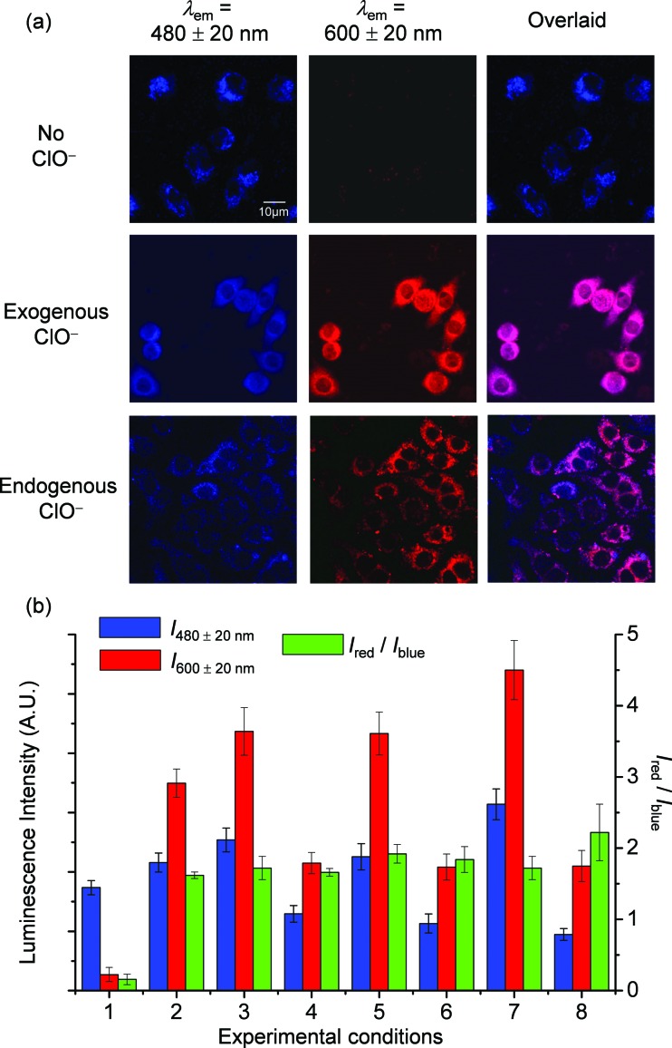
Fig. 6. (a) Luminescence images of RAW 264.7 cells treated with SiO2-1@mSiO2-2 (120 μg ml–1) for 3 h at 37 °C (top), followed by incubation with NaClO (50 μM) for 1 h (middle), and RAW 264.7 cells stimulated with LPS and PMA, and incubated with SiO2-1@mSiO2-2 (120 μg ml–1) for 2 h at 37 °C (bottom). (b) Luminescence intensity of RAW 264.7 cells recorded from the blue window (blue) and the red window (red) and the intensity ratio I red/I blue (green). Experimental conditions: RAW 264.7 cells were treated with SiO2-1@mSiO2-2 (120 μg ml–1) for 3 h at 37 °C without (1) and with (2) further incubation with NaClO (50 μM) for 1 h; the concentration of SiO2-1@mSiO2-2 was increased to 180 μg ml–1 (3) and decreased to 60 μg ml–1 (4); the incubation duration of SiO2-1@mSiO2-2 was increased to 6 h (5); the incubation temperature of SiO2-1@mSiO2-2 was reduced to 25 °C (6); the 405 nm laser power was randomly increased (7); and detection of endogenous ClO was recorded (8).

Fig. 6