Fig. 4.
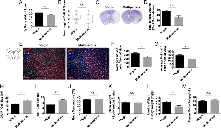
Reproductive experience attenuates brain injury following ischemic stroke. Virgin and multiparous mice were subject to sham or stroke surgical conditions and evaluated at 72 h to determine differences in histopathology and physiology. The percentage of body weight loss after stroke was smaller for multiparous females (A) (n = 12–15 per group). Clinical assessment shows that neurological deficit scores at 72 h approached but did not achieve significance between virgin and multiparous mice (B) (n = 12–15 per group). Total hemispheric infarct volume measurements were determined using cresyl violet-stained brain sections. Representative virgin and multiparous cresyl violet-stained ischemic brains (C) and quantification of infarct volumes (D) are shown (n = 10–12 per group). Representative immunohistochemistry of DAPI+ nucleated GFAP+ astrocytes and Iba1+ microglia/macrophages in the ischemic cortex (E). (Scale bar, 100 μm.) The average number of astrocytes (F) and microglia/macrophages (G) counted in the ischemic cortex is shown (n = 10–12 per group). The average cell size was quantified for astrocytes and microglia/macrophages (H and I, respectively), which were larger in multiparous mice. No differences in core body temperature (degrees Celsius) or normalized spleen weight were found between groups after stroke (J and K, respectively; n = 12–14 per group). Quantification of uterine weights (L) and plasma estradiol concentrations (M) at 72 h is shown (n = 12–14 per group). Error bars show mean SEM. DAPI, 4′,6-diamidino-2-phenylindole; GFAP, glial fibrillary acidic protein; Iba1, ionized calcium binding adaptor molecule 1; n.s., not significant; px, pixel. *P < 0.05; **P < 0.01; ***P < 0.001.

