Fig. 8.
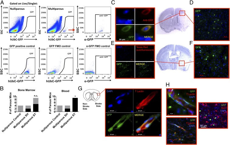
Fetal microchimeric cells were found in the maternal brain after ischemic injury. Low-frequency GFP+ fetal microchimeric cells were seen in maternal blood, bone marrow, and brain after ischemic stroke. Representative dot plots demonstrate the presence of GFP-positive fetal cells in maternal, but not nulliparous, bone marrow (A). The colabeling of endogenous GFP expression with anti-GFP+ fetal cells is consistent with the staining pattern found in transgenic GFP-expressing mice [A; positive and cell-specific fluorescence minus one (FMO) controls]. The red arrow refers to the GFP-positive cells in the Top Middle plot which were subsequently evaluated by SSC vs. anti-GFP in the Top Right plot. As shown in B, the frequency of parous females in which circulating GFP+ fetal cells (of total live CD45+) were detected was significantly increased at 24 h after stroke (SH = 0.007–0.008% vs. ST = 0.001–0.267%), whereas no change was found for those detected in bone marrow (SH = 0.002% vs. ST = 0.001%). Representative immunohistochemistry reveals clustering of GFP+ fetal cells in affected cortical (D) and striatal (F) brain regions across the anterior/posterior axis. (Magnification: D and F, 40×.) Validation of GFP specificity was proven using an anti-GFP antibody (C) and by the absence of autofluorescence (E). (Magnification: C, 100×; E, 20×.) Fetal cells persisted in the maternal brain for up to 30 d after stroke (G and H). At this later timepoint, GFP+ fetal cells adopted endothelial properties as evidenced by elongated morphology and tomato lectin staining (G and H). Our data shows that some fetal cells exhibit amoeboid morphology, but did not express markers associated with microglia/macrophages (H). Error bars show mean SEM. (Magnification: G, 100×; H, Top, 100×; H, Bottom and Right, 63×.) FM, fetal microchimerism; n.s., not significant; SSC, side light scatter. *P < 0.05.

