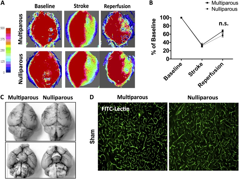
Fig. S2.
Baseline comparison of cerebral blood flow and vascular anatomy. Representative images of Laser speckle perfusion depict cerebral blood flow at baseline, during MCAO, and after reperfusion (A). Quantification of CBF flux demonstrated similar reductions in both groups (B) (n = 4 per group). There were no differences in large vessel anatomy, including the circle of Willis (C). The cortical microvasculature was assessed by i.v. injection of FITC-lectin using immunohistochemistry. Representative images showed no comparable differences in the staining pattern of FITC-lectin–labeled blood vessels (green) between nulliparous and multiparous mice (D). Error bars show mean SEM. (Magnification: A, 5×; D, 10×.) CBF, cerebral blood flow; FITC, fluorescein isothiocyanate; n.s., not significant.

