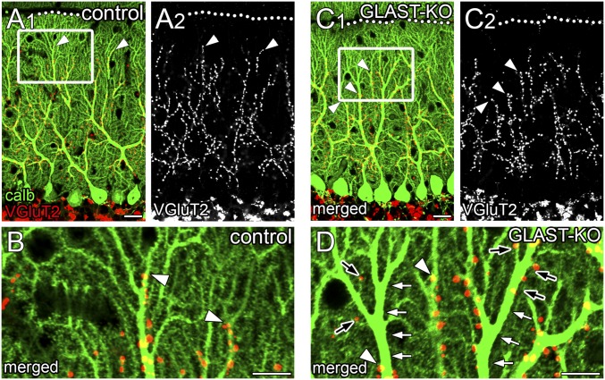
Fig. 2.
Perturbed dendritic innervation by ascending CF branches. Double immunofluorescence for calbindin (green) and VGluT2 (red or gray) in control (A and B) and mutant (C and D) mice. B and D are boxed regions in A1 and C1, respectively. Arrowheads indicate the distal tips of VGluT2(+) CF terminals. In mutant mice; dendritic shafts often lack CF innervation (white arrows, D), but a few CF terminals reappear on such vacant portions of proximal dendrites or emerge at distal dendrites (black arrows, D). Dotted lines indicate the pial surface. (Scale bars: A and C, 20 µm; B and D, 10 µm.)

