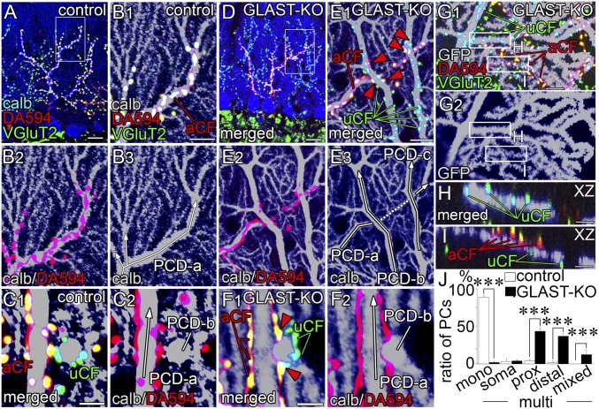
Fig. 3.
Multiple CF innervation at proximal (A–F) and distal (G–I) dendrites. (A–F) Triple labeling for calbindin (blue or gray), VGluT2 (green), and DA594 anterograde tracer (red) in control (A–C) and mutant (D–F) mice on parasagittal (A, B, D, and E) and horizontal (C and F) sections. B and E are enlarged images of boxed regions in A and D, respectively. Red arrowheads in E and F indicate aberrant innervation of tracer-labeled CFs that cause multiple CF innervation at proximal dendrites. White arrows indicate the course and branching of proximal shaft dendrites. (G–I) Triple labeling for GFP (gray), DA594 (red), and VGluT2 (green) in a GFP-transfected mutant PC, showing multiple innervation at distal dendrites. Boxed regions in G are enlarged as XZ-plane images of H and I. (J) Histogram showing the pattern and location of CF innervation: CF monoinnervation (mono) and multiple CF innervation (multi). The location of additional CF innervation is classified into somatic type (soma), proximal dendritic type (prox, >2 µm in caliber), distal dendritic type (distal, <2 µm), and mixed type (mixed). n = 189 cells from six control mice, n = 347 cells from eight mutant mice. ***P < 0.001, Mann–Whitney U test. Error bars represent SEM. aCF, VGluT2 (+)/anterograde tracer-labeled CF; PCD, Purkinje cell dendrite; uCF, VGluT2 (+)/tracer-unlabeled CF. (Scale bars: A and D, 20 µm; B, E, and G, 10 µm; C and F, 5 μm; H and I, 2 μm.)

