Fig. S1.
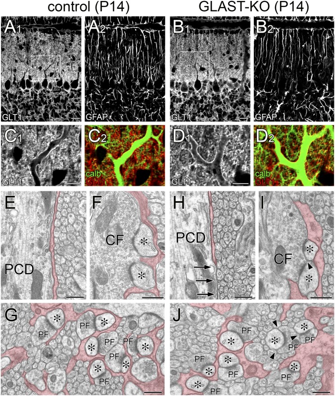
Mild impairment of BG coverage in GLAST-KO mice at P14. Light and electron microscopic images from control (A, C, and E–G) and mutant (B, D, and H–J) mice. (A–D) Triple immunofluorescence for glutamate transporter GLT-1 (A1–D1, C2, D2, red or gray), glial fibrillary acidic protein (GFAP; A2 and B2, gray), and calbindin (C2 and D2, green). Boxed regions in A and B are enlarged in C and D, respectively. No notable changes are discerned at the light microscopic level. In both mouse strains, shaft dendrites are similarly fringed with GLT-1-positive BG processes, and GFAP-positive Bergmann fibers are regularly arrayed in the molecular layer. (E–J) Electron micrographs of proximal shaft dendrites (E and H), CF–PC synapses (F and I), and PF–PC synapses (G and J). Arrows (H) indicate dendritic membrane without coverage of BG processes. Arrowheads (I and J) indicate the edges of the synaptic cleft without BG coverage, which contact with other neuronal elements. BG processes are pseudocolored in red. Asterisks indicate PC spines contacting CF or PF terminals. (Scale bars: A and B, 20 µm; C and D, 10 µm; E–J, 500 nm.)

