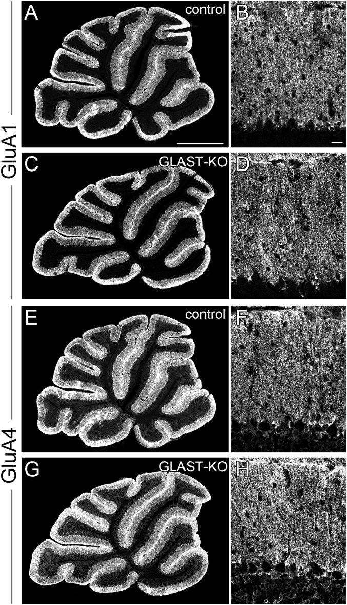
Fig. S2.
Immunofluorescence for AMPA receptor GluA1 (A–D) and GluA4 (E–H) subunits in control (A, B, E, and F) and GLAST-KO (C, D, G, and H) mice. In the molecular layer of the cerebellum, conventional immunofluorescence (without section pretreatment with pepsin) preferentially detects AMPA receptors on BG, whereas immunofluorescence with pepsin pretreatment unmasks synaptic AMPA receptors (33). In this analysis, we adopted conventional immunofluorescence to compare GluA1 and GluA4 expression on BG. In GLAST-KO mice, the pattern and intensity of GluA1 and GluA4 immunolabeling in the molecular layer are similar to those in control mice, suggesting GLAST-KO BG normally express GluA1 and GluA4. (Scale bars: A, 1 mm; B, 20 µm.)

