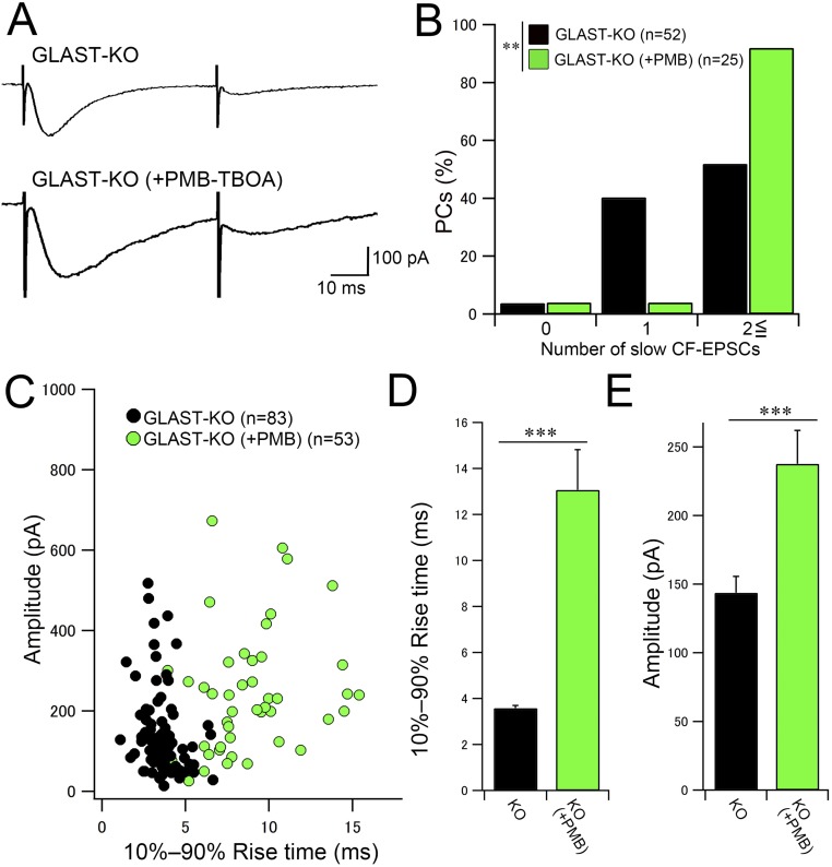
Fig. S7.
PMB-TBOA treatment significantly increases the number of slow CF-EPSCs in GLAST-KO mice. (A) Representative traces of slow CF-EPSCs in response to paired CF stimulation with an interpulse interval of 50 ms. Note that slow CF-EPSCs in 200 nM PMB-TBOA–treated mutant mice (Bottom) have slower kinetics than those in untreated mutant mice (Upper). Vh, −80 mV. (B) Frequency–distribution histogram showing the number of discrete CF-EPSC steps with a slow 10–90% rise time (>1 ms) in untreated (black columns) and PMB-TBOA–treated (green columns) mutant mice. Note that the probability of occurrence of slow CF-EPSCs is significantly increased by PMB-TBOA application in mutant mice. ***P < 0.001, Mann–Whitney U test. (C) Scatter plot of the amplitude (ordinate, pA) and the 10–90% rise time (ms, abscissa) of slow CF-EPSCs measured at a holding potential of −80 mV. (D and E) Summary bar graphs showing the average 10–90% rise time (D) and amplitude (E) of slow CF-EPSCs from untreated (black columns) and PMB-TBOA–treated (green columns) mutant mice. The data for mutant mice without PMB-TBOA treatment in B–E are the same as those shown in Fig. 4. Error bars represent SEM. ***P < 0.001; **P < 0.01; post hoc Dunn’s test. Numbers of PCs and responses examined are shown in parentheses. Holding potential was corrected for liquid junction potential.

